Advanced extractions in veterinary dentistry
Exodontia of mandibular and maxillary molars, maxillary fourth premolars, and canine teeth are frequently indicated in veterinary dentistry. Differences in the location and anatomy of each type of tooth present unique challenges, and require distinct surgical techniques. In this session, explore the anatomy of these teeth and their radiographic appearance. Review common indications for extraction as well as appropriate alternatives. Understand potential extraction complications—including orbital trauma mandibular fracture, and transportation of root fragments into the mandibular canal and nasal passages—and how to avoid them. You’ll also learn about a 3-corner mucoperiosteal flap technique that helps preserve the collar of attached gingiva surrounding neighboring teeth that are not being treated.
This session originally was presented at AVMA Convention 2023.
Learning Objectives:
- Learn about surgical techniques for extracting challenging teeth.
- Understand the anatomy and radiographic appearance of canines, molars, and maxillary fourth premolars.
- Understand flap design and handling.
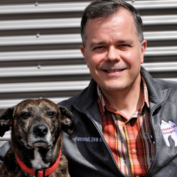
Dr. Patrick Vall earned his DVM from The Ohio State University. He spent the first 17 years of his career in general practice and emergency medicine, and he has owned practices in Maryland and Colorado. In 2015 he became board certified by the American Veterinary Dental College. In 2017 he became the owner of Animal Dental Care and Oral Surgery in Colorado. Dr. Vall is a past president of the Colorado Veterinary Medical Association Chapter 7-Colorado Springs Area Veterinary Society.